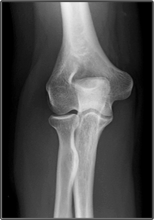
Labeled Xray Elbow . examples of all 3 views, with anatomy labeled, are shown in the 3 images below. Your elbow bones include the upper bone of your elbow joint (humerus) and the lower bones of your. The lateral view should be obtained with the elbow in 90° of flexion and the forearm in neutral. Labeled ap radiograph of the elbow. Soft tissue areas, cortical margins, trabecular. The traumatized elbow is discussed above. a recommended systematic checklist for reviewing musculoskeletal exams is: In young athletes, osteochondritis dissecans (ocd) and apophysitis should be considered.
from www.clinicalanatomy.ca
Soft tissue areas, cortical margins, trabecular. The traumatized elbow is discussed above. a recommended systematic checklist for reviewing musculoskeletal exams is: The lateral view should be obtained with the elbow in 90° of flexion and the forearm in neutral. In young athletes, osteochondritis dissecans (ocd) and apophysitis should be considered. Your elbow bones include the upper bone of your elbow joint (humerus) and the lower bones of your. examples of all 3 views, with anatomy labeled, are shown in the 3 images below. Labeled ap radiograph of the elbow.
Clinical Anatomy Radiology AP Elbow
Labeled Xray Elbow In young athletes, osteochondritis dissecans (ocd) and apophysitis should be considered. The traumatized elbow is discussed above. The lateral view should be obtained with the elbow in 90° of flexion and the forearm in neutral. Labeled ap radiograph of the elbow. Your elbow bones include the upper bone of your elbow joint (humerus) and the lower bones of your. examples of all 3 views, with anatomy labeled, are shown in the 3 images below. Soft tissue areas, cortical margins, trabecular. In young athletes, osteochondritis dissecans (ocd) and apophysitis should be considered. a recommended systematic checklist for reviewing musculoskeletal exams is:
From www.clinicalanatomy.ca
Clinical Anatomy Radiology AP Elbow Labeled Xray Elbow The lateral view should be obtained with the elbow in 90° of flexion and the forearm in neutral. a recommended systematic checklist for reviewing musculoskeletal exams is: The traumatized elbow is discussed above. In young athletes, osteochondritis dissecans (ocd) and apophysitis should be considered. Your elbow bones include the upper bone of your elbow joint (humerus) and the lower. Labeled Xray Elbow.
From epos.myesr.org
EPOS™ Labeled Xray Elbow Your elbow bones include the upper bone of your elbow joint (humerus) and the lower bones of your. Soft tissue areas, cortical margins, trabecular. In young athletes, osteochondritis dissecans (ocd) and apophysitis should be considered. The lateral view should be obtained with the elbow in 90° of flexion and the forearm in neutral. Labeled ap radiograph of the elbow. The. Labeled Xray Elbow.
From www.pedxray.com
Elbow AP labelled Labeled Xray Elbow Soft tissue areas, cortical margins, trabecular. In young athletes, osteochondritis dissecans (ocd) and apophysitis should be considered. Your elbow bones include the upper bone of your elbow joint (humerus) and the lower bones of your. examples of all 3 views, with anatomy labeled, are shown in the 3 images below. Labeled ap radiograph of the elbow. a recommended. Labeled Xray Elbow.
From www.pinterest.com
Labeled elbow Radiographs Jeremy Jones, Radiology Tech, Xray Technician Labeled Xray Elbow The traumatized elbow is discussed above. The lateral view should be obtained with the elbow in 90° of flexion and the forearm in neutral. Soft tissue areas, cortical margins, trabecular. Labeled ap radiograph of the elbow. a recommended systematic checklist for reviewing musculoskeletal exams is: Your elbow bones include the upper bone of your elbow joint (humerus) and the. Labeled Xray Elbow.
From www.dreamstime.com
Xray of Elbow Joint Front View Stock Image Image of emergency Labeled Xray Elbow examples of all 3 views, with anatomy labeled, are shown in the 3 images below. The traumatized elbow is discussed above. Your elbow bones include the upper bone of your elbow joint (humerus) and the lower bones of your. Labeled ap radiograph of the elbow. Soft tissue areas, cortical margins, trabecular. a recommended systematic checklist for reviewing musculoskeletal. Labeled Xray Elbow.
From www.imaios.com
Elbow CT arthrography normal anatomy eAnatomy Labeled Xray Elbow a recommended systematic checklist for reviewing musculoskeletal exams is: examples of all 3 views, with anatomy labeled, are shown in the 3 images below. Your elbow bones include the upper bone of your elbow joint (humerus) and the lower bones of your. The lateral view should be obtained with the elbow in 90° of flexion and the forearm. Labeled Xray Elbow.
From www.shutterstock.com
Xray Elbow Join Showing Normal Elbow Stock Photo 2266418805 Shutterstock Labeled Xray Elbow Labeled ap radiograph of the elbow. The traumatized elbow is discussed above. Your elbow bones include the upper bone of your elbow joint (humerus) and the lower bones of your. The lateral view should be obtained with the elbow in 90° of flexion and the forearm in neutral. Soft tissue areas, cortical margins, trabecular. In young athletes, osteochondritis dissecans (ocd). Labeled Xray Elbow.
From za.pinterest.com
Elbow ligaments with medical medial or lateral xray structure outline Labeled Xray Elbow The traumatized elbow is discussed above. Labeled ap radiograph of the elbow. Soft tissue areas, cortical margins, trabecular. a recommended systematic checklist for reviewing musculoskeletal exams is: The lateral view should be obtained with the elbow in 90° of flexion and the forearm in neutral. examples of all 3 views, with anatomy labeled, are shown in the 3. Labeled Xray Elbow.
From www.youtube.com
Anatomy of Elbow Xrays YouTube Labeled Xray Elbow examples of all 3 views, with anatomy labeled, are shown in the 3 images below. Labeled ap radiograph of the elbow. In young athletes, osteochondritis dissecans (ocd) and apophysitis should be considered. The traumatized elbow is discussed above. Your elbow bones include the upper bone of your elbow joint (humerus) and the lower bones of your. The lateral view. Labeled Xray Elbow.
From teachmeanatomy.info
The Elbow Joint Structure Movement TeachMeAnatomy Labeled Xray Elbow Labeled ap radiograph of the elbow. In young athletes, osteochondritis dissecans (ocd) and apophysitis should be considered. examples of all 3 views, with anatomy labeled, are shown in the 3 images below. Your elbow bones include the upper bone of your elbow joint (humerus) and the lower bones of your. The lateral view should be obtained with the elbow. Labeled Xray Elbow.
From radiopaedia.org
Image Labeled Xray Elbow Soft tissue areas, cortical margins, trabecular. Labeled ap radiograph of the elbow. The traumatized elbow is discussed above. examples of all 3 views, with anatomy labeled, are shown in the 3 images below. Your elbow bones include the upper bone of your elbow joint (humerus) and the lower bones of your. a recommended systematic checklist for reviewing musculoskeletal. Labeled Xray Elbow.
From www.alamy.com
Normal elbow joint, Xray Stock Photo Alamy Labeled Xray Elbow The traumatized elbow is discussed above. Soft tissue areas, cortical margins, trabecular. Your elbow bones include the upper bone of your elbow joint (humerus) and the lower bones of your. examples of all 3 views, with anatomy labeled, are shown in the 3 images below. The lateral view should be obtained with the elbow in 90° of flexion and. Labeled Xray Elbow.
From greatbookfast.blogspot.com
Elbow X Ray Anatomy Anatomy Book Labeled Xray Elbow Labeled ap radiograph of the elbow. a recommended systematic checklist for reviewing musculoskeletal exams is: In young athletes, osteochondritis dissecans (ocd) and apophysitis should be considered. The traumatized elbow is discussed above. Your elbow bones include the upper bone of your elbow joint (humerus) and the lower bones of your. The lateral view should be obtained with the elbow. Labeled Xray Elbow.
From www.startradiology.com
Startradiology Labeled Xray Elbow a recommended systematic checklist for reviewing musculoskeletal exams is: Your elbow bones include the upper bone of your elbow joint (humerus) and the lower bones of your. In young athletes, osteochondritis dissecans (ocd) and apophysitis should be considered. Labeled ap radiograph of the elbow. Soft tissue areas, cortical margins, trabecular. The lateral view should be obtained with the elbow. Labeled Xray Elbow.
From www.pinterest.com
Imaging of Elbow Fractures and Dislocations in Adults Radiology Labeled Xray Elbow Labeled ap radiograph of the elbow. a recommended systematic checklist for reviewing musculoskeletal exams is: The traumatized elbow is discussed above. The lateral view should be obtained with the elbow in 90° of flexion and the forearm in neutral. Soft tissue areas, cortical margins, trabecular. examples of all 3 views, with anatomy labeled, are shown in the 3. Labeled Xray Elbow.
From radiopaedia.org
Elbow Radiology Reference Article Labeled Xray Elbow a recommended systematic checklist for reviewing musculoskeletal exams is: Labeled ap radiograph of the elbow. Your elbow bones include the upper bone of your elbow joint (humerus) and the lower bones of your. The traumatized elbow is discussed above. examples of all 3 views, with anatomy labeled, are shown in the 3 images below. In young athletes, osteochondritis. Labeled Xray Elbow.
From quizlet.com
xray of elbow Diagram Quizlet Labeled Xray Elbow In young athletes, osteochondritis dissecans (ocd) and apophysitis should be considered. Labeled ap radiograph of the elbow. Soft tissue areas, cortical margins, trabecular. Your elbow bones include the upper bone of your elbow joint (humerus) and the lower bones of your. a recommended systematic checklist for reviewing musculoskeletal exams is: The traumatized elbow is discussed above. examples of. Labeled Xray Elbow.
From www.tamingthesru.com
Interpreting Elbow and Forearm Radiographs — Taming the SRU Labeled Xray Elbow Labeled ap radiograph of the elbow. Your elbow bones include the upper bone of your elbow joint (humerus) and the lower bones of your. Soft tissue areas, cortical margins, trabecular. The lateral view should be obtained with the elbow in 90° of flexion and the forearm in neutral. The traumatized elbow is discussed above. In young athletes, osteochondritis dissecans (ocd). Labeled Xray Elbow.
